 Home page of the UKE?
 DE
DE
Morphology and Electron Microscopy
/bilder/b%C3%BChnen/neu_3.png)
Our facility covers a large spectrum of light and electron microscopy techniques with a major focus on sample preparation and characterisation of genetically engineered animals, including immunolocalisation of proteins, ultrastructural analysis, and data processing.
We advice interested scientists on morphological questions and teach and train researchers in the application of microscopy techniques. Finally, our facility introduces and establishes new techniques and guarantees efficient use of the respective equipment.
DFG registered facility (RI00590)
Services
- Performance of light- and electron microscopical investigations
- Advice and practical instruction in the application of histochemical techniques
- Instruction of researchers in operation of microscopes and accessories
- Introduction of useful new (immuno-) histochemical techniques and/or equipment
Techniques
- Morphological studies of many kinds of tissues with light-, confocal laser scanning-, or transmission electron microscopy
- Patho-histological analysis of the whole body of transgenic mice
- Histo- (cyto) chemical staining procedures
- Immunohisto- (cyto) chemistry
- Pre- and postembedded immunogold labelling techniques (Tokuyasu)
- Correlated light-and electron microscopy (Tokuyasu CLEM)
- Negative staining of extracellular vesicles (EVs)
- Ultramicrotomy including serial sectioning
- Energy-dispersive X-ray spectroscopy (EDX)
We prepare cell and tissue samples for scientific histological and (immuno-) histochemical light and fluorescence microscopy. All preparation steps, (including fixation, sectioning with vibratome, cryotome or microtome, staining, mounting etc.) are performed by the group.
Volume EM and 3D imaging
- TEM tomography
- Serial section reconstruction
Equipment
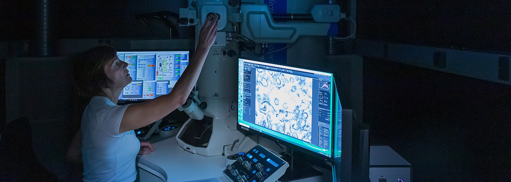
The Core Facility for Morphology and Electron Microscopy is equipped with two transmission electron microscopes.The electron microscopy laboratory including technical staff is available for sample preparation.
For section preparation we use two Leica EM Ultracut 7 and one Leica Ultracut S.
Electron Microscopes
TEM JEOL JEM 2100plus
TEM ZEISS 900
Regulations and usage costs
Contact
Dr. Imke Fiedler & PD Dr. Oliver Kretz, Co-Leaders
Imke Fiedler,
Oliver Kretz,
Chudamani Raithore,
Saskia Siegel,
Emanuela Szpotowicz,